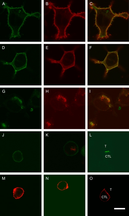
Figure 7. Slp2-a localizes predominantly to the plasma membrane and focuses at the immunological synapse.

Mouse Slp2-a tagged with pmaxGFP at either the amino terminus (panels A–C) or the carboxy terminus (panels D–I) expressed in RBL cells (panels A–I). Panels A, D and G show GFP fluorescence, and B and E show staining with anti-Slp2-a SHD antibody. Panel H shows Lgp100 staining. Panels C, F and I are merged images of A and B, D and E, and G and H, respectively. Panels J–O show N-terminally pmaxGFP-tagged mouse Slp2-a expressed in mouse CTL either unstained (panels J and L) or counterstained with anti-Lamp2 antibody ABL-93 (panel K) or anti-Slp2-a SHD antibody (panels M–O). Letter T indicates P815 target cell. All panels to the same scale. Scale bar: 10 μm.
