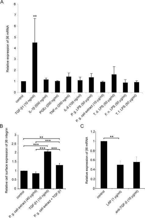
Figure 4.
P. gingivalis, LAP, and anti-TGF-β1 reduce αvβ6 integrin expression in cultured gingival keratinocytes. A: Gingival keratinocytes were treated with cytokines (TGF-β1, IL-1β, prostaglandin E2, TNF-α, IL-6), bacterial LPS (P. gingivalis, T. denticola, F. nucleatum, T. forsythensis), or P. gingivalis whole cell lysate. Total RNA was extracted, and the expression of β6 integrin subunit was analyzed by real-time PCR using β-actin as a control. PCR reactions were performed in triplicates, and the experiments were repeated four to seven times. B: The cells were exposed to cell lysate of P. gingivalis, TGF-β1, or a combination of both. Total cell surface expression of αvβ6 integrin was measured by flow cytometry. The experiment was repeated three times. C: The cells were treated with LAP or function-blocking anti-TGF-β1 antibody. Relative expression of β6 integrin was analyzed as in A. The experiment was repeated three times. Relative β6 integrin expression is expressed as mean ± SD of parallel experiments in all figures. Only differences that were deemed statistically significant are indicated: *P < 0.05; **P < 0.01, ***P < 0.001 (analysis of variance and Tukey’s post test or Student’s t-test). Results of the effect of anti-TGF-β1 antibody treatment are from a single experiment, and SD was calculated from three parallel samples.

