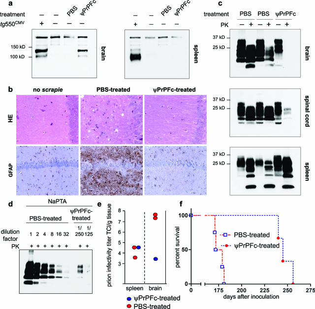
Figure 2.
Intracerebral lentiviral vector administration. a: Western blot analysis of frontal brain of transgenic PrP-Fc2 mice,8 and PBS-treated and ψPrPFc-treated animals. Using anti-PrP antibody,3 sustained PrP-Fc2 expression was detected 6 months after virus injection in the brain of ψPrPFc-treated animals, but not in the spleen. b: Severe spongiosis and astrogliosis in terminally sick PBS-treated animals, but no sign of prion-associated pathology in a ψPrPFc-treated animal sacrificed 170 dpi, similarly to age-match uninoculated mice. Sections were stained with H&E and immunostained with GFAP for detection of astrocytosis. c. Whereas PrPSc signals were visible in wt brains and spinal cords at terminal stage, no or weak signals were present in the brain and spinal cord of a ψPrPFc-treated animal sacrificed at the same time point. However, spleens of treated and controls animals exhibited similar quantities of PrPSc. d: NaPTA-enhanced quantitative Western analysis showed that PrPSc accumulation in the tested ψPrPFc-treated brain was 0.03% of the tested PBS-treated brain. To reach a comparable intensity level for Western blot quantification, control samples were diluted linearly up to 32 times, whereas for ψPrPFc-treated brains, PrPSc was concentrated by NaPTA precipitation from 125- or 250-fold more starting material than was used directly for Western blot in the case of PBS-treated mouse, lane 1. e: Prion infectivity, determined in the brain of one ψPrPFc-treated animal by the scrapie cell assay in endpoint format (SCEPA), was reduced by 4.2 log TCl/g tissue compared to PBS-treated animals. f: Survival of scrapie-infected mice treated with PBS or ψPrPFc. Whereas PBS-treated mice (n = 4) succumbed to disease at 175 ± 5 dpi, ψPrPFc-treated animals (n = 3) had an extended survival of 72 days (247 ± 8 dpi).

