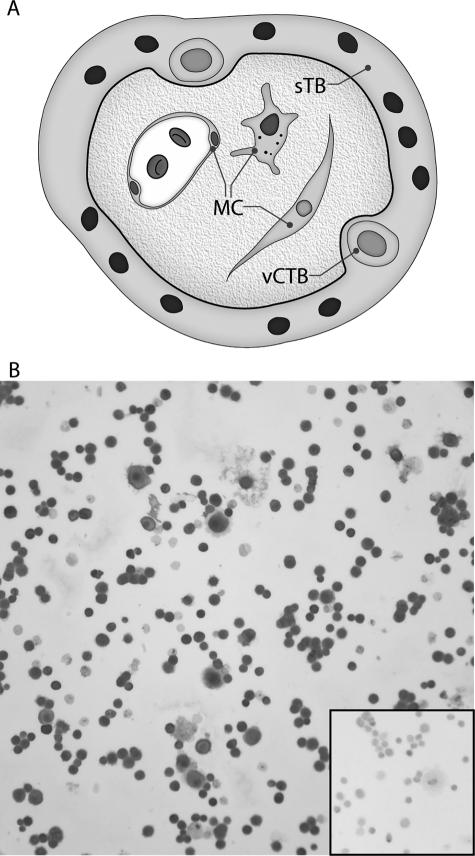
Figure 1.
Immunocytochemical identification of trophoblast cells in the vCTB cell preparations used in the experiments. A: Cartoon showing locations of vCTB and syncytiotrophoblast (sTB) cells and MCs in human term placental villus. B: Identification of cytokeratin-7-positive trophoblast cells. The percentage of trophoblast cells in the vCTB cell preparations was established by centrifuging the cells onto glass slides using a Shandon Cytospin (Shandon, Pittsburgh, PA) and immunostaining for cytokeratin-7 to reveal trophoblast cells. The cells were counterstained with Mayer’s hematoxylin. The inset shows failure of binding of the isotype control mAb. Original magnifications, ×200.

