Figure 1.
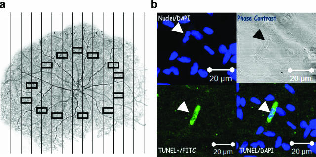
Diabetes increases retinal microvascular cell apoptosis. a: A low power image of a typical retinal trypsin digest of the retinal microvasculature is shown. Vertical lines represent the boundaries of contiguous fields in which the entire retina was examined for the presence of TUNEL- or cleaved caspase-3-positive cells. Rectangles in the mid-retinal area represent the areas examined for acellular capillaries and pericyte ghosts. Original magnification ×20. b: Representative image of a TUNEL-positive retinal microvascular cell in a diabetic specimen. Upper left panel, DAPI staining; lower left panel, TUNEL staining; upper right panel, phase contrast image of the corresponding field; lower right panel, merged TUNEL/DAPI images. Arrow points to a TUNEL-positive microvascular cells. The bar represents 20 μm at original magnification ×400.

