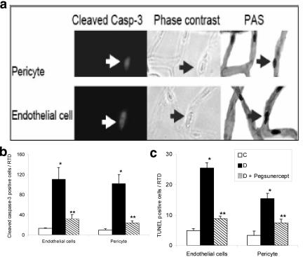
Figure 3.
TNF inhibition reduces pericyte and endothelial cell apoptosis in type 2 diabetic retina. a: Representative image of a cleaved caspase-3-positive pericyte (upper left panel) and endothelial cell (lower left panel), phase contrast (central panel), and PAS staining (right panel). Arrow points to a cleaved caspase-3-positive microvascular cells in a diabetic specimen. b: The mean number of cleaved caspase-3-positive endothelial cells and pericytes was determined in normoglycemic rats (C), Zucker diabetic fatty rats (DM) and Zucker diabetic fatty rats treated with pegsunercept (DM + Peg). Data represent the mean ± SEM (n = 5). c: The mean number of TUNEL-positive retinal endothelial cells and pericytes was determined in normoglycemic control rats (C), ZDF diabetic rats (DM), and ZDF diabetic rats treated with pegsunercept (DM + Peg). Data represent the mean ± SEM (n = 6). *Statistically significant compared to control (P < 0.05). **Statistically significant as compared with diabetic (P < 0.05).

