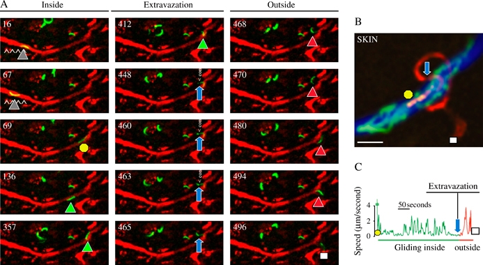
Figure 3. A Plasmodium berghei sporozoite exiting a blood vessel in the dermis of a mouse.

A) The fluorescent sporozoite glides inside the vessel colored in red after injection of red fluorescent BSA. The time (in seconds) is indicated in the upper left part of each panel. The intravascular sporozoite glides during the first 67 seconds (gray triangles), is suddenly displaced (yellow circle, 69 seconds), glides again inside the vessel (green triangles), extravazates (from 448 to 465 seconds, note the sporozoite constriction pointed by the blue arrows) before gliding in the dermis (468 to 496 seconds, red triangles until the white square). B) Maximum intensity projection of the fluorescent sporozoite from the 69th (yellow circle) to the 496th second (white square) through the constriction (blue arrow). C) Velocity profile of the sporozoite between the 69th and the 496th second.
