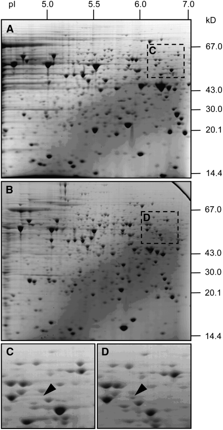
Figure 4.
Identification of a Cellulose Synthase Protein Fragment in Germinating Cysts with Appressoria.
(A) Two-dimensional gel showing proteins isolated from mycelia.
(B) Two-dimensional gel showing proteins isolated from germinating cysts with appressoria.
(C) Enlarged portion of gel in (A) showing location of the spot identified as Pi CesA1 from mycelia.
(D) Enlarged portion of gel in (B) showing location of the spot identified as Pi CesA1 from germinating cysts with appressoria.
Gels were equally loaded with 50 μg total protein and stained with Coomassie blue. Protein pI and mass (kD) are shown in (A) and (B) on the x and y axes, respectively. The spot identified as Pi CesA1 appears more abundant in gel (D) than in gel (C).

