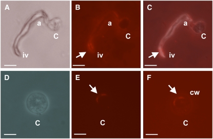
Figure 9.
CesA1-4 Proteins Localize to the Growing Tip and Infection-Like Vesicles of Appressoria and Cysts.
Wild-type P. infestans (strain 88069) cysts harvested from 11-d-old solid rye plates were induced to produce appressoria on plastic cover slips, overnight at 11°C. Cells were fixed and labeled with primary anti-CesA antibodies and visualized using Alexa Fluor 555. Phase contrast ([A] and [D]), fluorescent micrographs ([B] and [E]), and combined images ([C] and [F]) of a wild-type appressorium and a cyst. Arrows indicate areas of strong localization of CesA1-4 proteins. c, cyst; a, appressorium; iv, infection vesicle-like structure; cw, cell wall. Note that due to the fixing process used in this procedure, appressoria are dehydrated and therefore appear slightly deflated in (A) to (C). Bars = 10 μm in (A) to (C) and 5 μm in (D) to (F).

