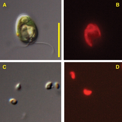
Figure 2.
C. reinhardtii and O. tauri.
(A) Differential interference contrast image of C. reinhardtii.
(B) Chlorophyll a fluorescence image of C. reinhardtii.
(C) Differential interference contrast image of O. tauri.
(D) Chlorophyll a fluorescence image of O. tauri.
The images were taken with a Zeiss Axioimager M1 fluorescence microscope. Total magnification for all images equals ×1250. Each image was captured from a different field of view, so the fluorescence images are not the same cells shown in the differential interference contrast images. Bar = 10 μm.

