
Figure 4.
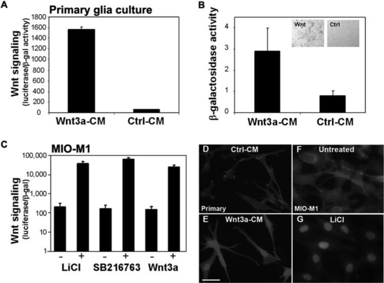
The Wnt signaling pathway is activated in Müller glia. (A) Addition of Wnt3a conditioned media (Wnt3a-CM) to primary Müller glia cultures increased activity of the Wnt luciferase reporter compared with Ctrl-CM. Luciferase activity is normalized to cotransfected β-galactosidase (values given in thousands). (B) Wnt3a also increased activity of β-galactosidase in Muller glia cultures from Tcf-LacZ retinas. B-gal activity is calculated per microgram of protein. Inset, blue: X-gal stain in Wnt3a-treated cultures (Wnt) and no stain in control treated cultures (Ctrl). (C) The MIO-M1 human Müller cell line activates Wnt signaling in response to Wnt3a ligand, LiCl, and SB216763 (+, treated with Wnt activator; –, control treatments (untreated for LiCl, DMSO for SB216763, control conditioned media for Wnt3a). A Wnt-responsive luciferase reporter was used to measure Wnt signaling in MIO-M1 cells as in (A). Log scale is shown. (A–C) Average ± SD. (D–G) Nuclear localization of β-catenin is used as an endogenous marker of Wnt signaling activation. Nuclear β-catenin is shown in primary Müller glia cultures (D, E) and the MIO-M1 Müller glia cell line (F, G) treated with the Wnt activators Wnt3a (E) and LiCl (G). The control for Wnt3a is Ctrl-CM (D) or untreated (F). Scale bar, 50 μm.