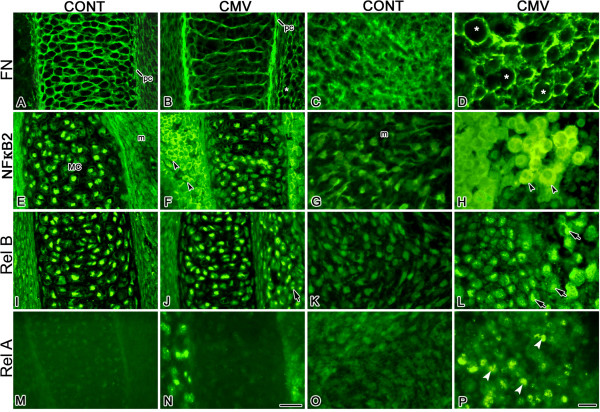
Figure 4.
mCMV infection altered the cell-specific localization of FN, NFκ-B2, RelB, and RelA proteins in E11 + 10 MANs. A-D. FN distribution. In controls (A, C), FN is normally seen on Meckel's cartilage chondroblasts, more weakly in the perichondrium (pc) and diffusely distributed throughout the ECM. With mCMV infection (B, D), FN is intensely localized in Meckel's cartilage perichondrium (pc) and more weakly on misaligned chondrocytes. Note that FN also surrounds individual cytomegalic mesenchymal stromal cells (*). E-H. NFκ-B2 distribution. In control (E) and mCMV-infected (F) MANs, nuclear-localized NFκ-B2 is seen in Meckel's cartilage chondrocytes; NFκ-B2 is absent from control mesenchymal (m) stromal cells. mCMV infection (F, H) induced de novo expression of cytoplasmically-localized NFκ-B2 in abnormal stromal cells (black arrowheads). I-L. RelB distribution. A similar RelB nuclear localization is found in Meckel's cartilage chondrocytes in control (I) and mCMV-infected (J) MANs. With viral infection (J, L), de novo expression of nuclear-localized RelB is seen in cytomegalic stromal cells (arrows). M-P. RelA distribution. RelA is not detected in Meckel's cartilages in control (M) and mCMV-infected (N) MANs; it is also absent from control mesenchymal stroma (M, O). mCMV-infected explants (N, P) exhibit de novo expression of nuclear-localized RelA (white arrowheads) in abnormal stromal cells. Bar, A-B, E-F, I-J, M-N: 30 μm; C-D, G-H, K-L, O-P: 10 μm.

