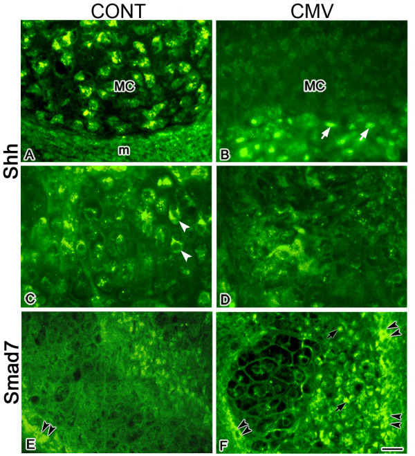
Figure 5.

mCMV infection induced a marked increase in immunodetectable Shh and Smad7 in E11 + 10 MANs. A, B. Shh expression in Meckel's cartilage. In controls (A), Shh protein is seen in Meckel's cartilage (MC) chondrocytes but not in the perichondrium or surrounding mesenchymal stroma (m). In contrast, mCMV-infected explants (B) exhibit de novo expression of Shh protein in abnormal, cytomegalic stromal cells (white arrows) surrounding Meckel's cartilage but is absent from Meckel's cartilage chondrocytes. C, D. Shh expression in mandibular bone. In controls (C), Shh is seen in mandibular bone (white arrowheads). In contrast, there is a marked decrease in Shh protein in mCMV-infected bone (D). E-F. Smad7 expression in mandibular bone. In controls (E), Smad7 is primarily localized in mandibular bone periostial cells (black double arrowheads). With mCMV infection (F), there is a substantial increase in immunodetectable Smad7 in mandibular osteoblasts (black arrows) and periostial cells (black arrowheads). Bar, A-F: 20 μm.
