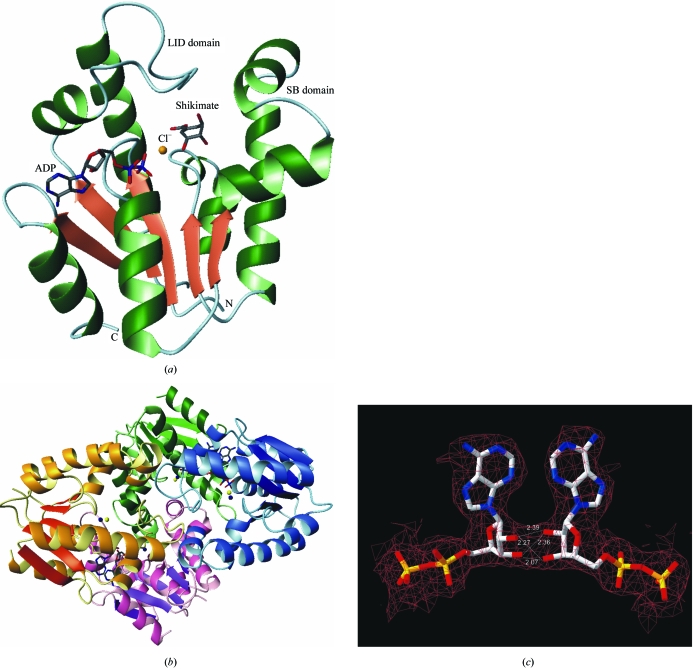
Figure 1.
(a) Structure of MtSK in complex with ADP, shikimate and Cl−. (b) Tetrameric structure of MtSK in complex with ADP, Mg2+ and Cl−. The monomers A, B, C and D are represented in green, blue, pink and yellow, respectively. The dark blue and yellow spheres represent the magnesium and chloride ions, respectively. The ADP molecules are represented as sticks. (c) Representation of the hydrogen-bonding interactions that occur between the ribose hydroxyl groups of ADP molecules bound to monomers A and B. The distances are shown in Å. Figures were generated with the program MolMol (Koradi et al., 1996 ▶).

