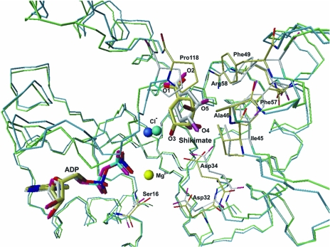
Figure 2.
Superposition of the structures of the MtSK–MgADP–shikimate and MtSK–ADP–shikimate ternary complexes. The Cα trace of the MtSK–MgADP–shikimate complex is presented in green and that of the MtSK–ADP–shikimate trace is presented in blue. The C atoms of MtSK–MgADP–shikimate and MtSK–ADP–shikimate are coloured white and yellow, respectively. The Mg2+ shown in yellow and the chloride ion shown in turquoise refer to the MtSK–MgADP–shikimate structure, while the chloride ion in dark blue refers to the MtSK–ADP–shikimate structure. The figure was generated with the program MolMol (Koradi et al., 1996 ▶).

