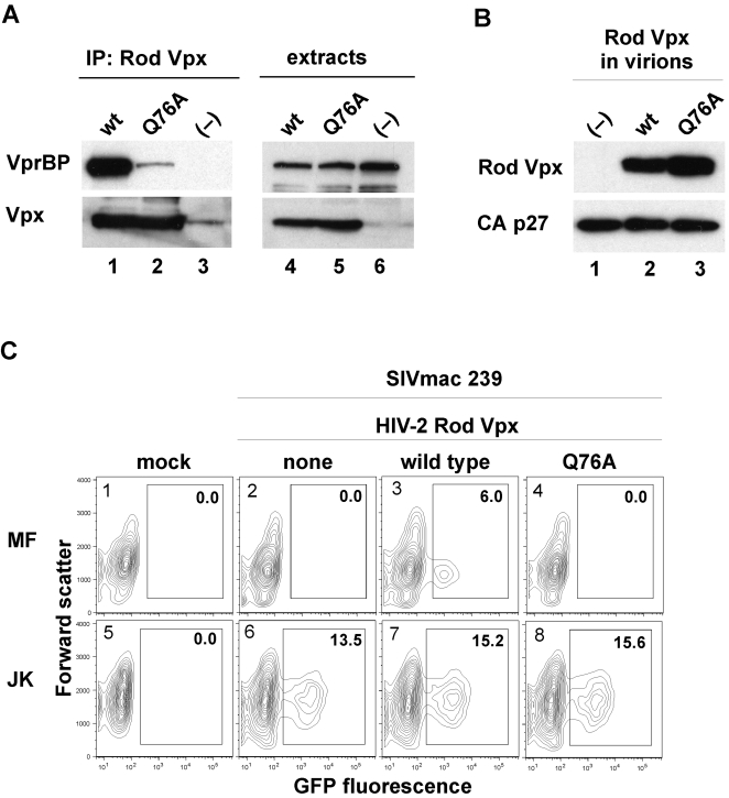
Figure 4. HIV-2 Rod Vpx facilitates macrophage transduction through its interaction with VprBP.
(A) HIV-2 Rod Vpx binds VprBP/DCAF1. hfa-tagged wild type (lanes 1 and 4) or Q76A substituted (lane 2 and 5) HIV-2 Rod Vpx proteins were transiently expressed in HEK 293T cells and precipitated from detergent extracts with FLAG-M2 affinity resin. VprBP and hfa-Vpx were detected in immune complexes (left panel) and cell extracts (right panel) by immunoblotting and visualized by enhanced chemiluminescence. (B) Wild type and Q76A substituted HIV-2 Rod Vpx proteins are incorporated into SIVmac virions. VSV-G pseudotyped single cycle SIVmac 239(GFP) virions were produced from HEK 293T cells transiently coexpressing wild type (lane 2) or Q76A mutated (lane 3) Rod Vpx, SIVmac 239(GFP) proviral clone with disrupted vpx and env genes, and VSV-G. Virions were partially purified and analyzed for Vpx and p27 Capsid by Western blotting. (C) Human monocyte-derived macrophages (MF, panels 1–4), and Jurkat T cells (JK, panels 5–8) were infected with normalized amounts of viruses characterized in (B) above, or mock infected (panels 1 and 5), and GFP positive cells were determined after 4 days (MF), or 2 days (JK), by flow cytometry. Percent fractions of GFP-positive cells (boxed) are indicated.

