
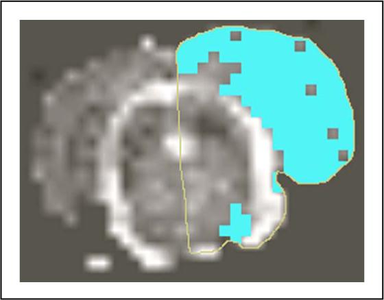
Figure 2.
Shows lesion size estimation from ADC map using MRVision software. Thresholding technique was used to extract lesion pixels on the ipsilateral hemisphere with ADC values < 80% of the contralateral side.
Official websites use .gov
A
.gov website belongs to an official
government organization in the United States.
Secure .gov websites use HTTPS
A lock (
) or https:// means you've safely
connected to the .gov website. Share sensitive
information only on official, secure websites.
Shows lesion size estimation from ADC map using MRVision software. Thresholding technique was used to extract lesion pixels on the ipsilateral hemisphere with ADC values < 80% of the contralateral side.