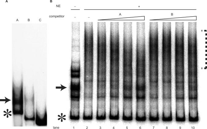
Figure 2.
Hoxb9 promoter fragment forms a secondary structure. (A) Native gel electrophoresis of fragments A, B and C revealed that only the fragment having promoter activity, fragment A, separated into multiple, but discrete, bands. Bands consisting of slow-moving secondary-structured DNA are indicated by arrows, and bands consisting of fast-moving linear DNA are indicated by asterisks. (B) EMSA performed in the absence (−) or presence (+) of nuclear extracts (NE) from P19 cells and competition assay for protein–DNA binding. Because a protein in the NE specifically binds the highly structured DNA fragment (indicated by arrow), the slow-moving band disappeared and formed a smear (indicated by dotted line) in the presence of NE, as shown in lanes 1 and 2. Lanes 3–10 show results from competition assays. The concentrations of competitor DNA used were 12.5-, 25-, 50- and 100-fold molar ratio relative to the probe (lanes 3 and 7; 4 and8; 5 and 9; 6 and 10, respectively). Adding fragment A as a competitor caused the band (arrow) to reappear (lanes 3–6). Fragment B as a competitor, however, was much less effective in causing the band to reappear, indicating that fragment B failed to compete successfully.

