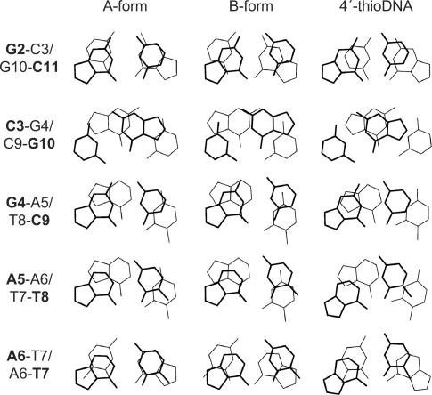
Figure 5.
The stacking pattern observed for fully modified 4′-thioDNA (right), with those for the canonical A-form (left) and B-form (middle), respectively, the terminal part being excluded. The views are perpendicular to base pairs, not along the helix axes. The upper bases are indicated by bold lines and characters.

