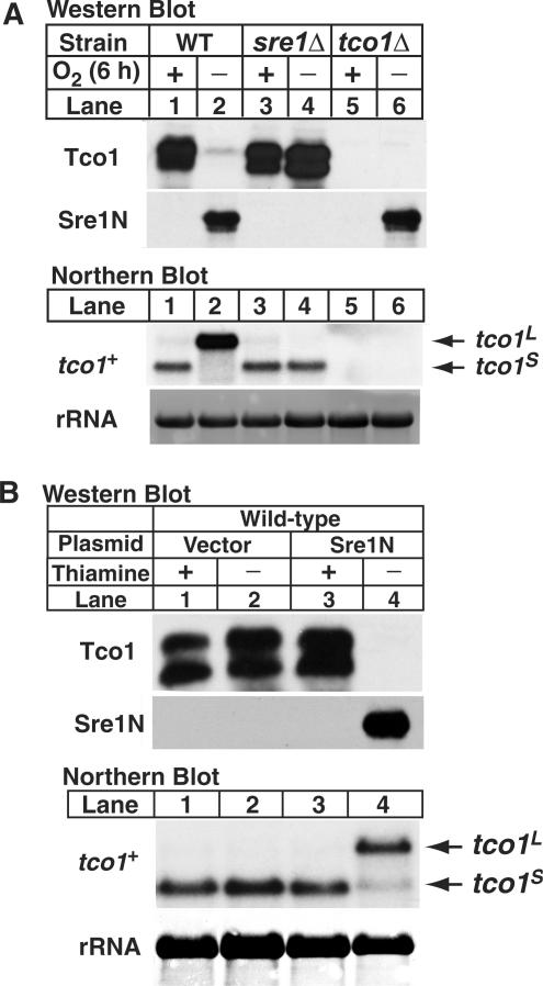
Figure 3.
Sre1 inhibits Tco1 protein expression. (A) Wild-type, sre1Δ, and tco1Δ cells were cultured for 6 h +/− oxygen. Upper panels: membrane protein samples (40 µg) were analyzed using antibodies raised against the C-terminus of Tco1 or cell lysates (40 µg) were analyzed by immunoblotting for nuclear Sre1. Lower panels: total RNA (10 µg) was subjected to northern analysis using a tco1+ probe. 25S rRNA was imaged as loading control. tco1L and tco1S denote the long and short mRNAs for tco1+, respectively. (B) Wild-type cells expressing sre1N from a plasmid under control of the thiamine repressible, nmt promoter were cultured in minimal medium in the presence (repressed) or absence (induced) of thiamine (5 µg/ml) for 24 h. Cells were diluted, cultured under the same conditions for 24 h and harvested in exponential phase. Upper panels: membrane proteins (36 µg) and cell lysates (40 µg) were immunoblotted using anti-Tco1 and anti-Sre1, respectively. Lower panels: total RNA (10 µg) was subjected to northern analysis using a tco1+ probe. 25S rRNA was imaged as loading control. tco1L and tco1S denote the long and short mRNAs for tco1+, respectively.

