Figure 3.
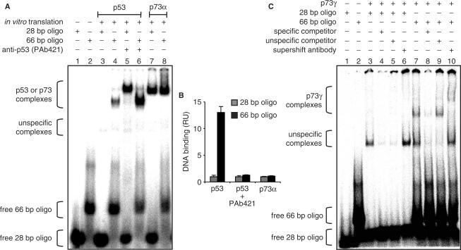
Enhanced binding of p53 and p73γ but not p73α to longer DNA oligonucleotides. (A) EMSA showing binding of in vitro translated p53 and p73α to double-stranded 32P labeled oligonucleotides containing 28 bp or 66 bp from the 5′ p53-binding site of the human p21 promoter. The p53 CTD was blocked by addition of 200 ng anti-p53 (PAb421) antibody. Protein–DNA complexes were visualized and quantified by phosphorimaging. (B) Graphical analysis of the phosphorimaging data depicted in (A). Binding was normalized to the 28 bp oligonucleotide. Data represent the mean ± SD of three independent EMSA experiments. RU, relative units. (C) DNA binding of in vitro translated p73γ as described in (A). A 200-fold excess of the same oligonucleotide without 32P label was used as a specific competitor, a scrambled sequence as an unspecific competitor.

