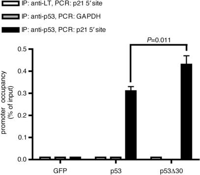
Figure 6.
Deletion of the p53 CTD enhances in vivo binding to the p21 promoter. ChIP (chromatin immunoprecipitation assay) comparing in vivo binding of p53 and p53Δ30 to the 5′ p53-binding site in the p21 promoter. Binding to a region within the GAPDH promoter is shown as a negative control. Binding to the p21 promoter of a control immunoprecipitation using an antibody directed against large T antigen (LT) is shown as an additional negative control. ChIP data were quantitated by qPCR and normalized to their respective inputs for p21 and GAPDH. The graph shows the mean and SD of promoter occupancy in percent of input DNA (n=3). p21 promoter occupancy of p53 and p53Δ30 were significantly different (P=0.011). Protein levels of p53 and p53Δ30 are shown in Figure 7B.

