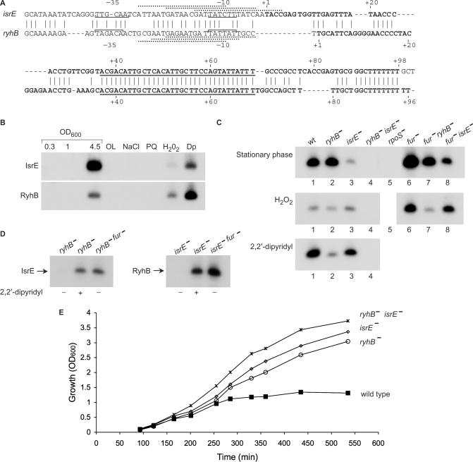
Figure 3.
Differential expression of isrE and ryhB. (A) Sequence of isrE and ryhB genes. The start sites observed by primer extension are indicated (+1). Brackets indicate putative −10 and −35 regions of the promoters mapped. The core sequences (nucleotides 37–69 relative to the start sites) are underlined. Three possible Fur binding sites that match Fur consensus (gataatgataatcattatc) at 15, 16 and 13 of 19 bases are indicated by dashed lines above the sequence of IsrE. Fur sites at ryhB, matching 14 of 19 bases of the consensus are indicated under the sequence of ryhB. (B) Detection of IsrE and RyhB in wild type cells. Northern blots as in Figure 2 of RyhB and IsrE using RyhB and IsrE specific primers (1271 and 797, respectively). The conditions used were; rich media (LB; at OD600 values of 0.3, 1 and 4.5, respectively), oxygen limitation (OL), osmotic shock (NaCl; 0.5 M NaCl), oxidative stress (PQ; 0.2 mM paraquat or H2O2; 1 mM hydrogen peroxide) and low iron conditions (Dp; 0.2 mM 2,2′-dipyridyl). (C) Late stationary phase cultures of wild type and mutants of isrE, ryhB, fur and rpoS were analyzed for isrE and ryhB levels by primer extension (top panel). Exponentially growing cultures were exposed to H2O2 (1 mM for 30 min. middle panel) or to 2,2′-dipyridyl (0.2 mM for 30 min. bottom panel). The primer extension reactions (0.5 μg RNA) were carried out using a primer (470) that is complementary to the core sequence of isrE and ryhB. (D) Fur regulation of isrE and ryhB as detected by primer extension. Exponentially growing cells were treated as in C. (E) Growth curves. Overnight cultures of wild type S. typhimurium and isrE and/or ryhB mutants were diluted 1/100 in LB medium and incubated shaking at 37°C. At 30 min after dilution the cultures were treated with 0.2 mM 2,2′-dipyridyl. Optical density was measured at the indicated time intervals.

