
Figure 2.
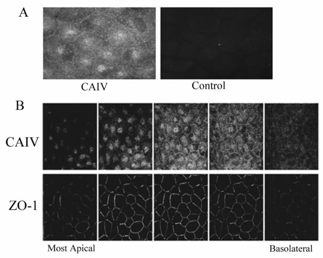
Immunofluorescence localization of CAIV in bovine corneal endothelium. A. fresh corneal endothelium; left, positive surface staining; right, control-absence of primary antibody. B. Confocal montage of cultured corneal endothelial cells stained for CAIV and ZO-1. Leftmost image pair is the most apical section. Z-axis separation between images is 0.5 μm. The five images represent a distance of 2.5 μm. CAIV fluorescence is either apical to or coincident with ZO-1.