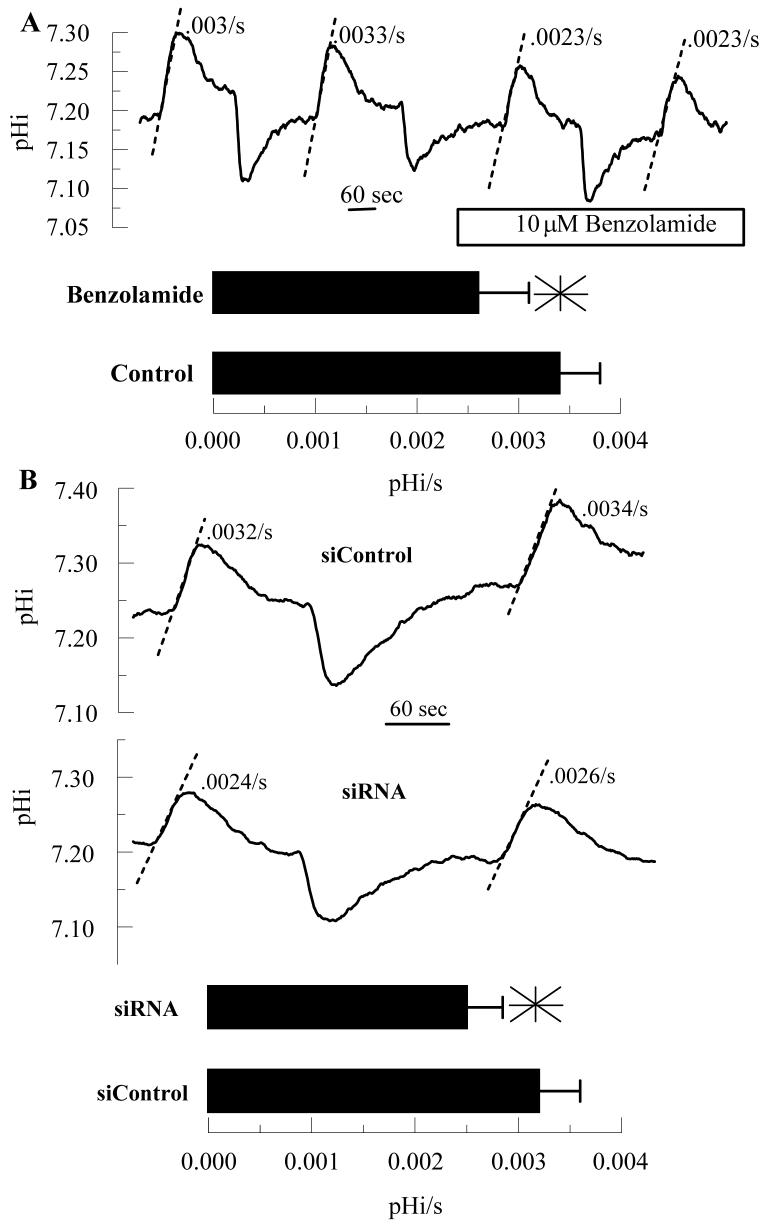
Figure 4.
Effect of Benzolamide and CAIV siRNA on Apparent CO2 fluxes. BCECF loaded endothelial cells were perfused in a two-sided chamber. The alkalinizations were due to changing the apical perfusate to a CO2/HCO3- free ringer. The dashed lines illustrate the estimated rate (pHi/s). A. This was performed twice in the absence and then in the presence of 10 μM benzolamide on the apical side. Bar graph shows mean rates and SD (n=12 anodiscs); *significantly lower than control (p<0.05, paired t-test). B. Representative comparisons of siControl and CAIV treated cells. Bar graph shows mean rates and SD (n=10 anodiscs); *significantly lower than control (p<0.05, indendent t-test).

