
Figure 6. Overexpression of β-arrestin2 enhances ligand-induced sequestration of FPR from the cell surface.
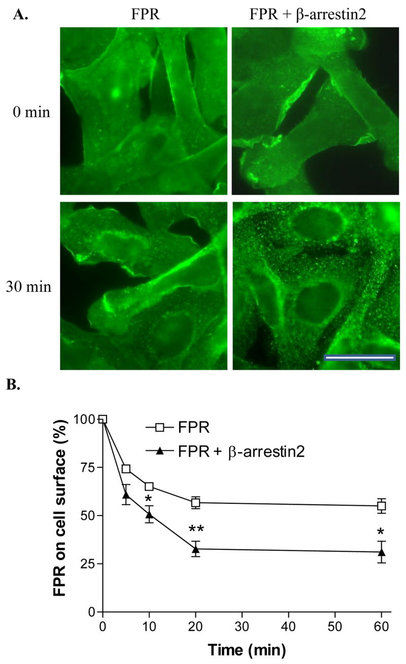
(A) CHO FPR and CHO FPR + β-arrestin2 cells were incubated for 0 or 30 min in the presence of 1 μM fMLF and subsequently stained with a polyclonal anti-FPR antibody. Bar 50 μm. (B) CHO FPR and CHO FPR + β-arrestin2 cells were incubated for 0, 5, 10, 20 or 60 min in the presence of 100 nM fMLF, washed in low pH buffer and the receptors remaining on the cell surface were detected by flow cytometry after 45 min binding of fluorescent ligand on ice. Graph shows means ± SD from three separate experiments. * P < 0.05; **P < 0.01.