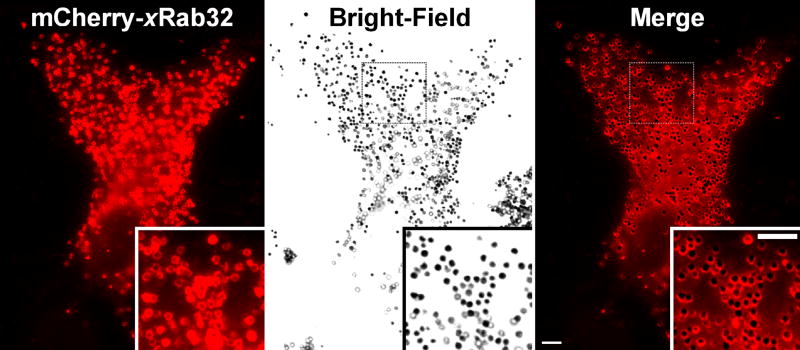
Figure 2. Rab32 is Localized to Melanosomes.
mCherry-xRab32 is localized to melanosomes. Xenopus melanophores were transfected with mCherry-xRab32 for 24 hr. The left panel shows the distribution of the mCherry-xRab32 fusion protein, the middle panel shows in bright-field distribution of melanosomes, and the right panel shows the bright field image merged with the distribution of mCherry-xRab32 fusion proteins. Melanosomes are decorated with mCherry-xRab32 (right panel inset), indicating that the mCherry-xRab32 fusion protein is localized to melanosomes. Bars, 5 μm.

