Figure 3.
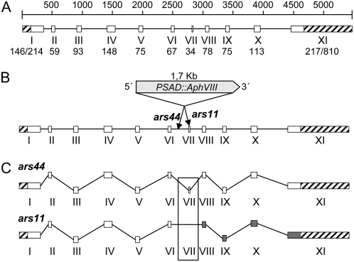
A, Genomic structure of SNRK2.1. The white blocks and lines represent exons and introns, respectively. Exons are numbered with roman numerals, and the corresponding sizes (bp) are given below. For exons I and XI, the two sizes given represent the 5′ UTR/CDS and CDS/3′ UTR, respectively. 5′ and 3′ UTRs are represented with striped blocks. The diagram is drawn to scale. B, Marker insertion in SNRK2.1 of ars11 and ars44. The PSAD∷AphVIII marker gene is inserted within exon VII in the ars11 mutant and within intron 6 in the ars44 mutant. In both cases, the marker gene has the same orientation as SNRK2.1. C, mRNA maturation in the ars11 and ars44 mutants. In the ars44 mutant, all of the introns appear to be properly spliced and a normal protein is synthesized. The ars11 mRNA lacks exon VII and an aberrant protein is synthesized. The gray blocks represent exons for which the reading frame has changed.

