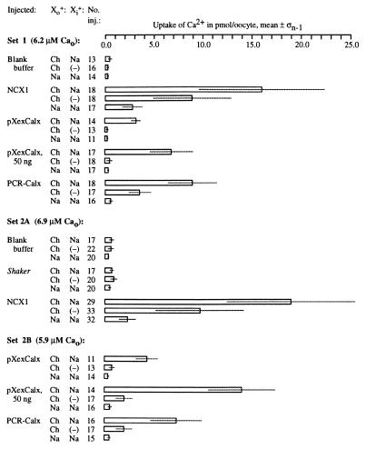
Figure 10.
Expression of CALX in Xenopus oocytes. Sets of oocytes were microinjected, incubated for several days, loaded with intracellular sodium, challenged with 45Ca2+ along with a variable monovalent cation, and allowed to take up 45Ca2+ via reverse Na-Ca exchange for 10′. The concentration of 45Ca2+ used (5.9–6.9 μM) is indicated for each oocyte set. After uptake, oocytes were rinsed and individually assayed by scintillation counting. “Injected” refers to the substance injected; Xo+, the primary monovalent cation placed outside the oocytes being assayed for 45Ca2+ uptake; Xi+, the primary monovalent cation loaded into the oocytes; “No. inj.,” the number of oocytes injected with a particular solution; “Uptake of Ca2+ in pmol/oocyte, mean ± σn−1,” the mean quantity and SD of 45Ca2+ taken up per oocyte. “Ch” denotes cationic choline; (−) denotes oocytes left unloaded by outside cations.

