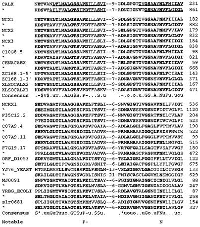
Figure 7.
Sequence alignment of Calx-α. CALX sequences predicted to be membrane-spanning are underlined. Each instance of a motif is shown with: its protein’s name; the motif’s sequence, with aligned regions separated by indicated numbers of unaligned residues; and the location of the motif’s C terminus in the protein. Proteins here are described in Fig. 3. All aligned regions have P values of <10−100 (24). Residues that match a consensus residue or residue type (shown in the Consensus line) are in boldface type. Residue types are defined as: aromatic (F, W, Y, or H, denoted @); aliphatic (I, L, or V, denoted u); acidic (D or E, −); basic (H, K, or R, +); charged (acidic or basic, ∗); hydroxylic (S or T, $), methyl (A, S, or C, μ), or small (G, A, S, or C, denoted o). A “⋅” denotes sites with no particular consensus. Note that the Calx-α motifs of CALX orthologs vs. paralogs have slightly different consensuses. Calx-α residues in the “Notable” line are noted in the Discussion.

