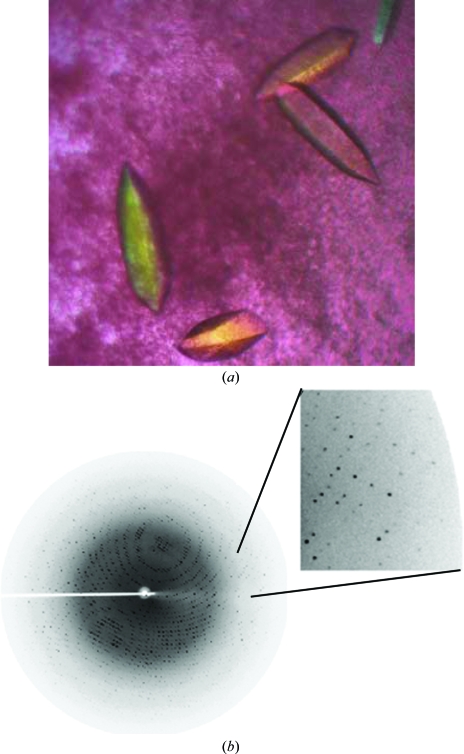
Figure 2.
(a) Single elongated hexagonal shaped crystals of EhCS obtained along with protein precipitate at 289 K using the hanging-drop vapour-diffusion method. (b) Diffraction pattern of EhCS crystals to 1.86 Å resolution. The data were collected using a Rigaku MicroMax-007 generator and a MAR imaging plate. The imaging plate was adjusted to a distance of 150 mm and the crystals were exposed for 90 s per frame. Diffraction spots were observed to the edges of the image plate, as shown in the enlargement.

