
Figure 9.
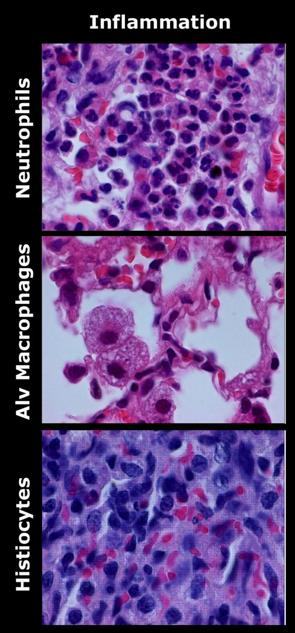
Histological examples of the inflammatory pattern of LIRI on HE slides (100*). Histological analysis confirms the flowcytometric analysis with the presence of predominantly neutrophils on day 1, alveolar macrophages on day 3, and histiocytes on day 30 following LIRI. HE = Haematoxylin and Eosin staining; LIRI = Lung Ischemia-Reperfusion Injury