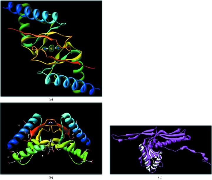
Figure 2.
(a) A ribbon diagram of the PF0899 dimer viewed down its twofold axis. The molecules are colored blue to red according to their sequence. The bridging dicyanoaurate ion with the Au atom sitting on the twofold axis is highlighted in the center. The dicyanoaurate is indicated as distinct atoms (Au, orange; C, gray; N, blue). Electron density (F o − F c OMIT map) for the dicyanoaurate ion is also shown contoured at 3σ. Images were generated with CHIMERA (Pettersen et al., 2004 ▶; http://www.cgl.ucsf.edu/chimera). (b) A ribbon diagram of the PF0899 dimer viewed perpendicular to its twofold axis. The positions of the three EXXE motifs are highlighted and labeled A, B and C. Note the large site C binding pocket located at the dimer interface. The color scheme is the same as in Fig. 2 ▶(a). Images were generated with CHIMERA. (c) Diagram showing the alignment and overlap of 1ohg and 2pk8.

