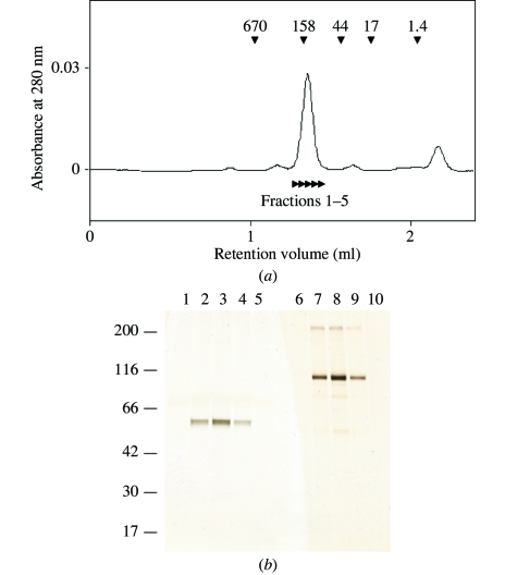
Figure 1.
(a) Size-exclusion chromatography of mGluR7 LBD. The elution positions of the marker proteins are shown by arrowheads with molecular weights in kDa. The estimated molecular weight of mGluR7 LBD is 157 kDa. The indicated fractions, 1–5, were subjected to SDS–PAGE analysis as shown in Fig. 1 ▶(b). (b) Aliquots of the peaks from the gel-filtration analysis were analyzed by SDS–PAGE. Samples were run in the presence (lanes 1–5) and absence (lanes 6–10) of DTT. The gel was stained with silver nitrate. The sizes of the molecular-weight markers are shown in kDa on the left. Lanes 1 and 6, fraction 1; lanes 2 and 7, fraction 2; lanes 3 and 8, fraction 3; lanes 4 and 9, fraction 4; lanes 5 and 10, fraction 5.

