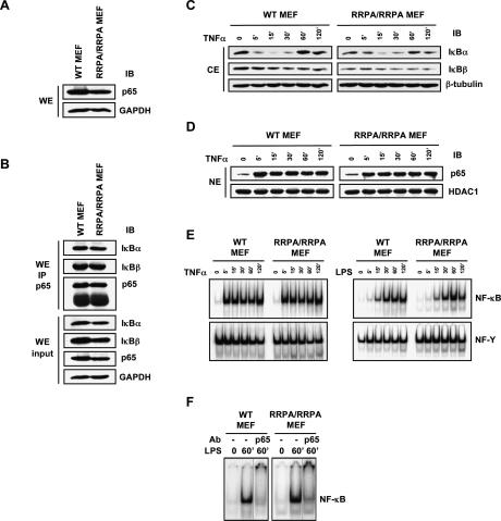
Figure 2.
Characterization of RRPA knock-in cells. (A) Immunoblot analysis of p65 expression levels in wild-type and RRPA homozygous MEFs generated from embryos at 12.5 dpc. (B) Interaction between p65 and IκBα or IκBβ in MEFs detected by immunoprecipitation. (C) Degradation and resynthesis of IκBα and IκBβ proteins in MEFs following TNFα stimulation (10 ng/mL). (D) p65 translocation in MEFs following TNFα stimulation. (E) EMSA using labeled κB probe and control NF-Y probe. Nuclear extracts (5 μg) from unstimulated MEFs and MEFs stimulated with TNFα or LPS (10 μg/mL) were analyzed. (F) Supershift analysis of NF-κB–DNA-binding complexes was performed using anti-p65 antibody. Nuclear extracts (5 μg) from unstimulated MEFs and MEFs stimulated with LPS for 60 min were analyzed.

