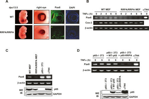
Figure 4.
Expression of Pax6 gene was diminished by RRPA mutant protein. (A) Pax6 expression in the eye. Embryonic eye cross-sections (13.5 dpc) were immunostained with the antibody recognizing Pax6 and then detected with secondary antibody coupled to Alexa 488. Nuclei were stained by DAPI. (B) Pax6 expression in untreated MEFs and in MEFs treated with TNFα. Gene expression levels were detected by semiquantitative RT–PCR. Cox-2 was used as a positive control for TNFα stimulation. αTN4 cells are mouse lens epithelial cells in which Pax6 is highly expressed, and were used as a positive control for Pax6 PCR analysis. (C) Pax6 was expressed at a high level in p65-deficient 3T3 cells detected by semiquantitative RT–PCR. p65 expression levels detected by immunoblotting are displayed in the bottom panels. (D) Pax6 expression was inhibited by RRPA mutant protein in stable cell lines. Wild-type p65 or p65-RRPA mutant was stably introduced into p65-deficient 3T3 cells. Pax6 levels were detected by semiquantitative RT–PCR. p65 expression levels detected by immunoblotting are displayed in the bottom panels.

