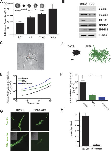
Figure 6.
Fn matrix assembly regulates the generation of 3D-specific myosin-dependent tension and intracellular rigidity. (A) 3D fibrin gels were cast in individual wells of 24-well plates and cultured alone or in the presence of embedded endothelial cells for 2 d in the presence of control IgG, mAb L8, mAb 9D2, the 70-kDa Fn fragment, or the FUD peptide. Gels were detached from the edges of the culture wells and contraction-monitored after an additional incubation period of 10 h at 37°C. The percentage inhibition of gel contraction (measured as the change in gel diameter) relative to an IgG control is presented. Inset shows photographs (from left to right) of a cell-free fibrin gel, a gel contracted by embedded endothelial cells, and gels contracted by endothelial cells cultured in the presence of either mAb 9D2 or mAb L8 or the 70-kDa Fn fragment. (B) Endothelial cells were cultured in 3D fibrin gels for 2 d with either the FUD peptide or the control Del29 peptide. Levels of β-actin, α-actinin, NMMIIA, NMMIIB, and MLC2 were measured by Western blot, with ERK1/2 serving as the loading control. As assessed by semiquantitative densitometry, the levels of β-actin, actinin, and MLC2 were 58 ± 7% (n = 5; mean ± 1 SD), 62% (n = 2), and 60 ± 12% (n = 3; mean ± 1 SD) of control. (C) Prior to embedding in the 3D fibrin matrix, endothelial cells were ballistically microinjected with 100-nm polystyrene beads. After 3-d incubation, the beads dispersed in the cytoplasm and their centroids were tracked with high spatial and temporal resolution using fluorescence microscopy. Bar, 10 μm. (D) Typical trajectories of beads in the cytoplasm of 3D-embedded cells, under control conditions (black, left panel) and in the presence of FUD (green, right panel). Time of movie capture, 20 sec. Bar, 50 nm. (E) Ensemble-averaged MSDs of beads in the cytoplasm of 3D-embedded endothelial cells under control conditions (black curve) and in the presence of FUD (green curve) or blebbistatin (blue curve). MSDs were computed from displacements of the beads such as shown in D. At least 110 beads in at least 10 cells were tracked for each condition. (F) Averaged elastic modulus of cells under control conditions or in the presence of FUD or blebbistatin. (***) P < 0.001. (G) Endothelial cells were cultured in 3D fibrin gels for 2 d in the presence or absence of 50 μM ± blebbistatin when F-actin organization and Fn matrix assembly were monitored by confocal laser microscopy. (Top panels) F-actin organization was monitored by staining with Alexa 488-conjugated phalloidin. (Bottom panels) Fn matrix assembly was assessed by culture in the presence of FITC-conjugated Fn. Bar, 20 μm. (H) Endothelial cells were cultured in 3D fibrin gels for 6 d under provasculogenic conditions in the presence of 50 μM ± blebbistatin or vehicle (DMSO). Tubulogenesis was monitored following sectioning, H&E staining, and lumen quantification.

