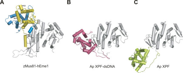
Figure 2.
Structural comparison between cMUS81ΔN and Ap XPF. Overall structures of the cMUS81ΔN complex (A), the Ap XPF-dsDNA complex (B), and apo Ap XPF (C) are shown. The nuclease domains (gray) from three structures are in the same orientation, and the rest of the structures including the HhH2 domains of each structure are colored yellow and blue (zMus81ΔN and hEme1ΔN), pink (Ap XPF-dsDNA) and green (apo Ap XPF). Equivalent secondary structures among three structures are numbered from H9 to H13.

