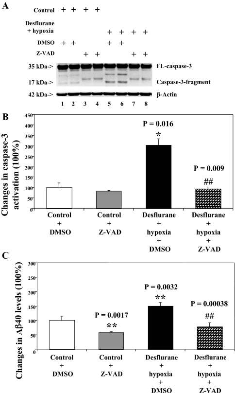
FIGURE 3.
Caspase inhibitor Z-VAD attenuates caspase-3 activation induced by desflurane/hypoxia in H4-APP cells. A, treatment with desflurane (12%) plus hypoxia (18%) for 6 h (lanes 5 and 6) induces caspase-3 cleavage (activation) as compared with control conditions (lanes 1 and 2) or Z-VAD (50 μm) treatment (lanes 3 and 4). Z-VAD treatment (lane 7 and 8) attenuates caspase-3 cleavage induced by desflurane/hypoxia. There is no significant difference in amounts of β-actin in H4-APP cells with above treatments. B, quantification of the Western blot shows that desflurane/hypoxia (black bar) increases caspase-3 activation as compared with control conditions (white bar) (*, p = 0.016), normalized to β-actin levels. The desflurane/hypoxia-induced caspase-3 activation is reduced by Z-VAD (50 μm) treatment (net bar; ##, p = 0.009). C, desflurane/hypoxia (black bar) increases secreted Aβ levels in H4-APP cells as compared with control conditions (white bar) (**, p = 0.0032). Z-VAD (net bar) reduces the desflurane/hypoxia-induced increases in secreted Aβ levels in H4-APP cells (##, p = 0.00038). DMSO, dimethyl sulfoxide.

