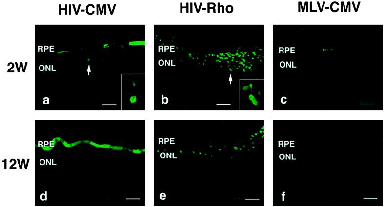
Figure 5.
Expression of GFP in the adult retina 2 and 12 weeks after injection. (a and d) HIV vector with the CMV promoter. (b and e) HIV vector with the rhodopsin promoter. (c and f) MLV vector with the CMV promoter. Arrows in a and b indicate GFP-positive photoreceptor cells. Insets in a and b, higher magnification of GFP-positive photoreceptor cells. Original magnification, ×400. Bars = 50 μm.

