
Figure 4. Increases in crypt heights are reduced in Klf5+/− mice following infection with C. rodentium.
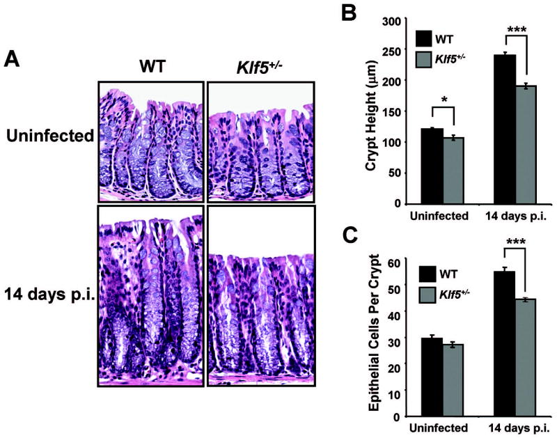
(A) H&E staining of paraffin-embedded distal colon tissues from WT C57BL/6 and Klf5+/− mice either uninfected (PBS controls) or infected with C. rodentium and sacrificed at 14 days p.i. (B) Crypt heights were determined from H & E stained sections measuring well-oriented crypts. Results represent the mean value of six mice, with the value of each mouse being determined from three separate crypt measurements. *P < 0.05, ***P < 0.001; n = 6. (C) Epithelial cells per crypt were counted from colonic crypts of H&E stained sections. Results represent the mean value of six mice, with the value of each mouse being determined by averaging cell counts from five crypts. ***P < 0.001; n = 6. See Supplemental Figures 3A and 3B, respectively, for scatter plots of the individual data values for Figures 4B and 4C.