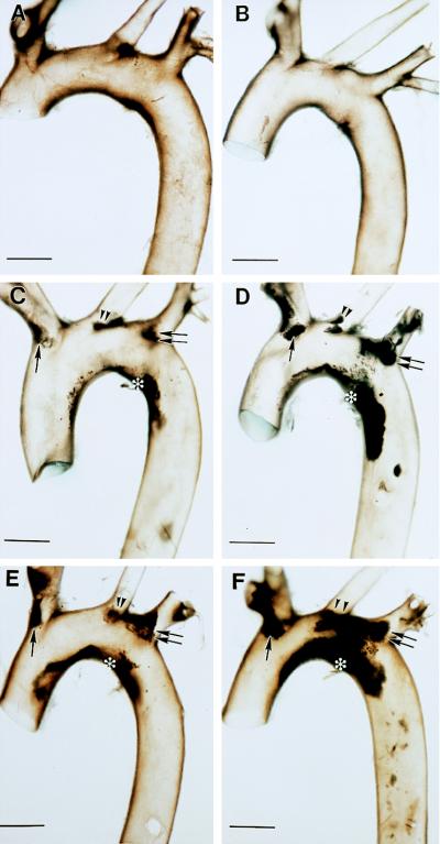
Figure 1.
Gross appearance of aortic arches of ApoE- and Plg-deficient mice. Brightfield view of the aortic arches collected from 26-week-old control (A) and Plg−/− (B) mice. Comparable views of typical aortic arches collected from 22-week-old (C and D) and 25-week-old (E and F), ApoE−/− (C and E) and ApoE−/−/Plg−/− (D and F) mice. Note the dark-appearing atherosclerotic lesions along the lesser curvature (asterisk), brachiocephalic trunk (single arrow), left common carotid (double arrowheads), and left subclavian (double arrows). Under darkfield, the lesions appear as opaque, cream-colored deposits. (Bars = 1 mm.)

