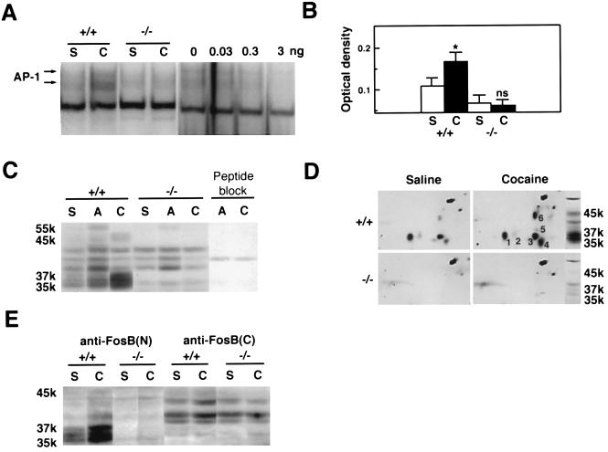
Figure 1.
Induction of AP-1 binding activity and FRAs in mouse striatum by cocaine. (A) Gel mobility shift assay with a consensus AP-1 sequence. S, repeated saline treatment; C, repeated cocaine treatment. +/+, wild-type littermates; −/−, fosB mutant mice. (Right) The autoradiogram demonstrates specificity of the AP-1 binding activity, by showing competition of the activity in striatum from a cocaine-treated wild-type mouse with a nonradioactive AP-1 probe (0, 0.03, 0.3, or 3 ng). (B) Levels of AP-1 activity. ∗, P < 0.05, statistically significant difference between saline- and cocaine-treated groups; ns, not statistically different. S+/+, n = 11; C+/+, n = 10; S−/−, n = 10; C−/−, n = 11. (C) Western blotting with the anti-FRA antiserum (ref. 4, see Materials and Methods). S, saline; A, acute (2 hr) cocaine treatment; C, repeated cocaine treatment. Peptide block, preadsorption of the FRA antiserum with the M-peptide immunogen (4) eliminated FRA bands. The band at ≈40 kDa is a nonspecific band. (D) Two-dimensional Western blotting with the anti-FRA antiserum. (E) Western blotting with antibodies directed against the N terminus of FosB [anti-FosB(N), which recognizes ΔFosB and FosB] or the C terminus of FosB [anti-FosB(C), which recognizes FosB only].

