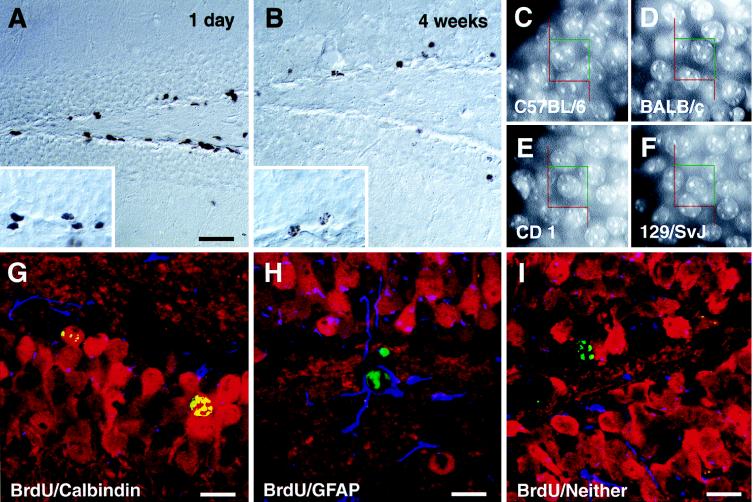
Figure 1.
Proliferation (A) and survival (B) of cells in the subgranular zone of C57BL/6 mice. BrdU-labeled cells 1 day after the last injection of BrdU (A) have dark irregular shaped nuclei (see Inset). Four weeks later (B) the number of BrdU-positive cells has decreased and the remaining cells have more rounded nuclei, sometimes with the typical chromatin structure of granule cells (see also insert and compare with C–F). Absolute granule cell numbers were determined stereologically using a 15 × 15 μm counting frame superimposed on a video image of Hoechst-33342-stained sections (C–F). No differences in the appearance of the granule cells can be noted, and the neuronal density in the granule cell layer is similar (see Table 2). Phenotypes of surviving newborn cells 4 weeks after the last injection were examined by means of immunofluorescence and confocal microscopy. Cells were categorized as to whether they showed double-labeling for BrdU (green) and granule cell marker calbindin (red) (G), BrdU and astrocytic marker GFAP (blue) (H), or for BrdU and neither of the two other markers (I). Quantification of phenotype distribution is found in Fig. 2B. (Bar in A = 75 μm for A and B and 25 μm for the Insets in A and B.) Counting frames in C–F are 15 × 15 μm. (Bars in G–I = 15 μm.)

