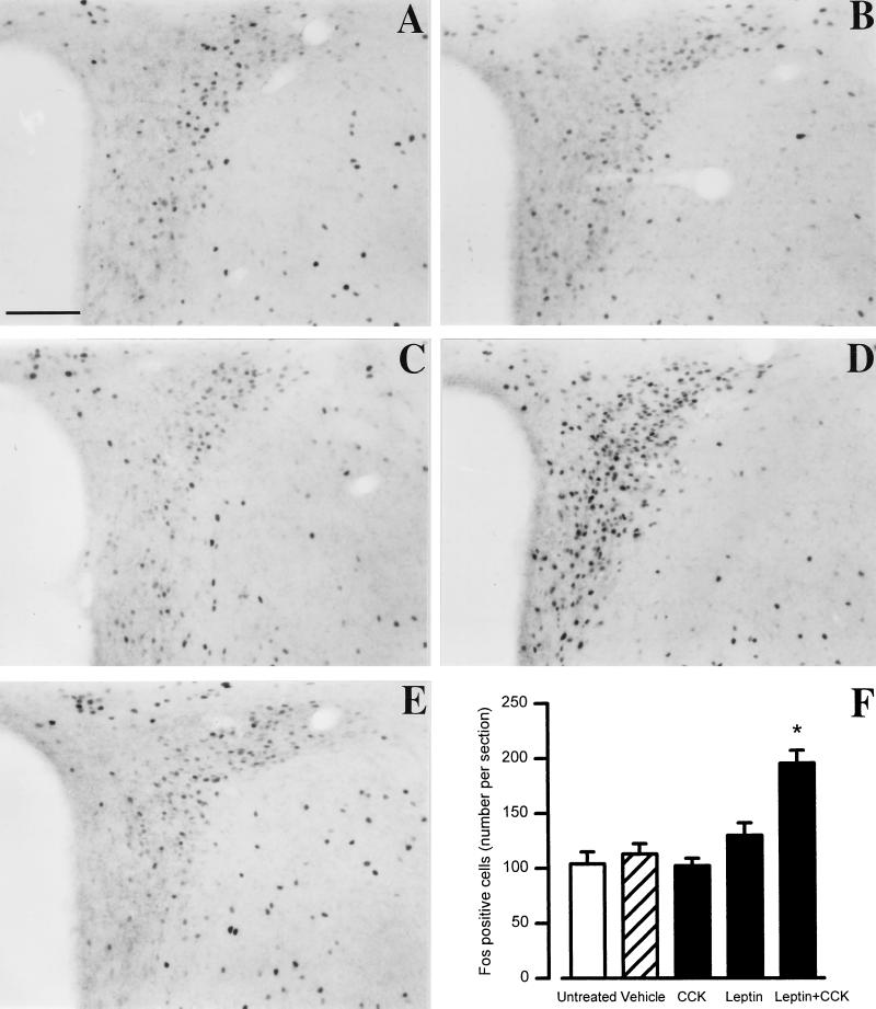
Figure 5.
Fos immunoreactivity in the PVN after leptin + CCK coinjected in lean mice (+/+). Representative microphotographs show Fos protein immunoreactivity in the PVN 2 hr after a single i.p. injection of (A) combined vehicles (10 ml/kg), (B) CCK (3.5 μg/kg), (C) leptin (120 μg/kg), or (D) leptin plus CCK, and (E) in fasted untreated animals. Marked increase in Fos immunoreactivity, resulting in dark and well-shaped nuclei, was observed in the parvo- and magnocellular divisions of the PVN after leptin plus CCK treatment, whereas other groups exhibited only scattered and lightly labeled cells. (Bar = 100 μm.) (F) The number of cells per section (unilateral). Data are mean ± SEM of four animals per group. ∗, P < 0.05 vs. all other groups (Kruskal–Wallis ANOVA, KW = −1308.4).

