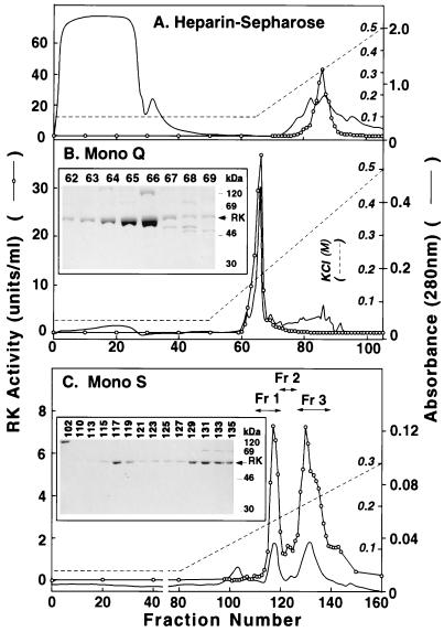
Figure 2.
Chromatographic Steps in Purification of RK. (A) Heparin-Sepharose. The crude extract (50 ml) prepared from 5 × 10⁸ infected cells was applied to a HiTrap-Heparin column and elution was performed. A280 (solid line) (scale on the outside) and RK activity (○) (scale inside A) are shown. (B) FPLC on Mono Q. Fractions 83–88 from A above (12 ml) were diluted and applied on Mono Q and elution was performed with a linear salt gradient. (Inset) Fractions 62–69 as examined by SDS/PAGE. (C) Fractions 63–66 (8 ml) from B above were chromatographed by FPLC on Mono S. RK activity was assayed. The three fractions indicated were pooled. (Inset). SDS/PAGE analysis of fractions 102–135.

