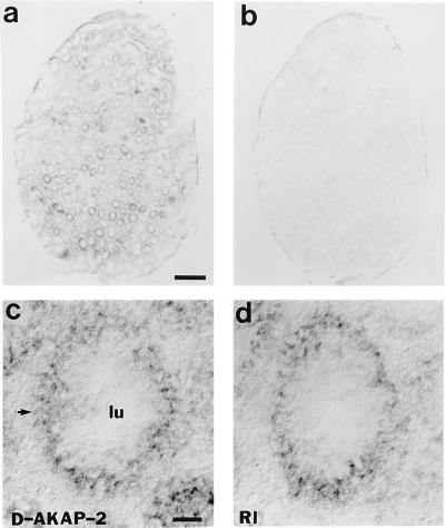
Figure 7.
In situ hybridization pattern of D-AKAP2 that shows overlapping regions for RIα. RNA probes of D-AKAP2 were derived from the cDNA sequence of 1–1469. RIα probes were derived from cDNA coding for residues 18–169. Hybridization was carried out on adjacent 20-μm cryostat sections of adult mouse testis and signals were visualized by alkaline phosphatase histochemistry. (a) D-AKAP1 mRNA is present in various regions of testis. (b) Signal of the sense strand as negative control. (c) High magnification of signal for D-AKAP2 on one of the testicular tube cross sections. (d) High magnification of signal for RIα on one of the testicular tube cross sections. (Bar = 100 μm.)

