
Figure 4. Prolonged exposure to a high concentration of DTDP abolishes force production in EDL and soleus fibres.
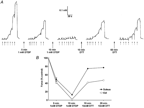
A, maximum force production in an EDL fibre was reduced to ≈40 % after a 5 min exposure to 1 mm DTDP (at pCa > 9), with the reduction in Ca2+ sensitivity being little different from that with brief (15 s) exposure to 100 μm DTDP (see text). Exposure to 1 mm DTDP for a further 10 min almost completely abolished force production, and force recovered to only ≈55 % of the original control level after a total of 70 min in DTT (pCa > 9). In each sequence the fibre was exposed successively to solutions at pCa 6.7, 6.41, 6.20, 6.00, 5.89, 5.75, 4.5, at times indicated by the small upward arrows. B, mean size (± s.e.m.) of maximum force in 3 soleus (♦) and 4 EDL (○) fibres after exposure to 1 mm DTDP for 5 min and then a further 10 min, and then to 10 mm DTT for two 10 min periods. Values are expressed relative to maximum force before exposure to DTDP. The s.e.m. is smaller than the symbol in most cases, showing that the effects are highly reproducible within each fibre type.