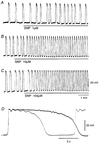
Figure 1. Effects of sodium nitroprusside (SNP) on pacemaker potentials recorded from the guinea pig antrum.

SNP (A, 1 μm; B, 10 μm; C, 100 μm) was applied as indicated by the bar under each record. D, high-speed trace of the pacemaker potential recorded in the absence (continuous line) and presence of 100 μm SNP (dotted line). The resting membrane potentials were: A, −63 mV; B, −62 mV; C and D, −64 mV. All responses were recorded from different tissues.
