
Figure 1. Technique to simultaneously stretch a guinea-pig ventricular myocyte and record electrical activity.
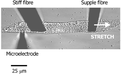
The microelectrode is used to record membrane potential or currents. It is placed behind the stiff fibre to protect the site of impalement during stretch. The supple fibre is used to stretch the cell and its displacement during stimulation is a measure of active force development (increased resting force caused by the stretch cannot be measured in this configuration). Increased sarcomeric spacing is used as the index of stretch.